X-rays: Health through Images
Radiography with X-rays is the starting point for diagnosing a variety of dental related issues and concerns. With the constant improvement of digital X-rays, offices can provide comfortable and painless care to our patients. Dental X-rays are taken to check the health of the patient’s mouth, gums and teeth. X-rays are simply pictures of the inside of your mouth, which can be used to diagnose problems such as cavities, tooth decay, infections and impacted teeth.
History of X-rays
Professor Wilhelm Conrad Roentgen first discovered the idea of X-rays in 1895. X-raying showed great potential for diagnosing medical concerns by showing the internal conditions of the body. Drotto Walkhoff took the first dental X-ray shortly after Roentgen’s discovery. As we know today, long term exposure to radiation is not very safe. Walkhoff discovered this himself as his constant exposure caused him to lose hair. As technology improved, X-ray images were taken at a faster rate and with less radiation. In the 1900’s, William Herbert Rollins drew up a list of precautions to better help minimize risks associated with X-ray exposure. His precautions also included enclosing the X-ray in lead housing to control the directions of the exposure as well as a lead apron to reject rays as X-rays are taken. Rollins also determined safe recommendations for exposure dosages on the average patient.
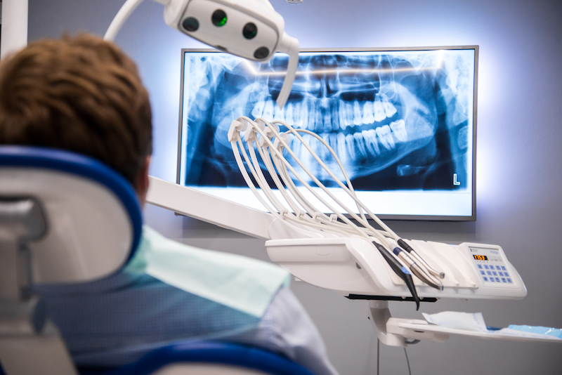
Digital X-rays today
Digital X-rays combine the power of computer technology with electronic sensors followed by very small bursts of radiation. Instead of processing the film, the images form as soon as the sensor is placed in the patient’s mouth and projected to the computer screen. Many offices these days only offer digital X-rays for patients. Digital dental X-rays are more cost efficient, quicker in speed and development, cleaner for the patient and staff and much safer than traditional X-rays.
- Cost effective: Because offices have switched to digital, they only use one digital sensor for all images verses using all individual single use films to create a full set of X-rays. Over time, the film cost will add up.
- Faster results: Traditional X-rays must go through a process to be developed, which takes up plenty of time in a time restricted appointment. With digital X-rays, there is no wait! As soon as the picture is taken with the sensor, it is up and ready to be viewed by the doctor.
- Better quality: Traditional X-rays are very limited on the number of shades used in the image. Digital X-rays can allow 250+ variations of greys. The computer also allows multiple options to enhance the X-rays for better viewing and diagnosing.
- Exposure levels: The most important benefit of digital over traditional X-rays is the safety. Digital X-rays can reduce radiation exposure for the patient up to 90%. In fact, you get more radiation on a cross country flight than you do taking digital X-rays!
Radiography has made significant leaps and bounds over the recent years and is now even bigger than when it was first discovered. As newer technology develops, X-raying in the medical and dental field will only improve making it simpler for the technicians and easier on the patients. Here at Smile Sarasota, we are always up to date on digital technology and the improvements made through our programs to make our dental visits with our patient safe and secure.
References:
https://columbiasurgery.org/news/2015/09/17/history-medicine-dr-roentgen-s-accidental-x-rays